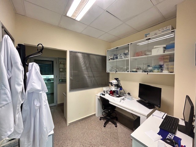
Research & Facilities
Click any research area below to explore our full capabilities, methods, and image galleries. From mouse preimplantation embryo development to advanced molecular biology, Yucar Lab provides a fully equipped research environment for groundbreaking discoveries in reproductive health.
Mouse Preimplantation Embryo Development
We investigate mouse preimplantation embryo development as a model system to understand the cellular and molecular mechanisms governing the earliest stages of mammalian life. Our research focuses on the transition from the zygote to the blastocyst stage, with particular emphasis on embryonic morphogenesis, lineage specification, trophectoderm and inner cell mass differentiation, cell–cell communication, and mechanisms underlying developmental arrest. By analyzing key regulatory molecules, including c-Abl, YAP/p73, TEAD4, CDX2, NANOG, PARD6, and E-cadherin, we aim to define the signaling networks that coordinate early embryonic patterning and cell fate decisions. These studies provide mechanistic insight into embryo quality, early developmental competence, and the molecular basis of infertility-related developmental failure.
- —Advanced mouse model systems
- —Regulatory mechanism analysis
- —In vitro fertilization and embryo culture
- —Single-cell gene expression profiling

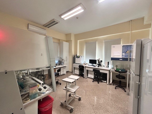
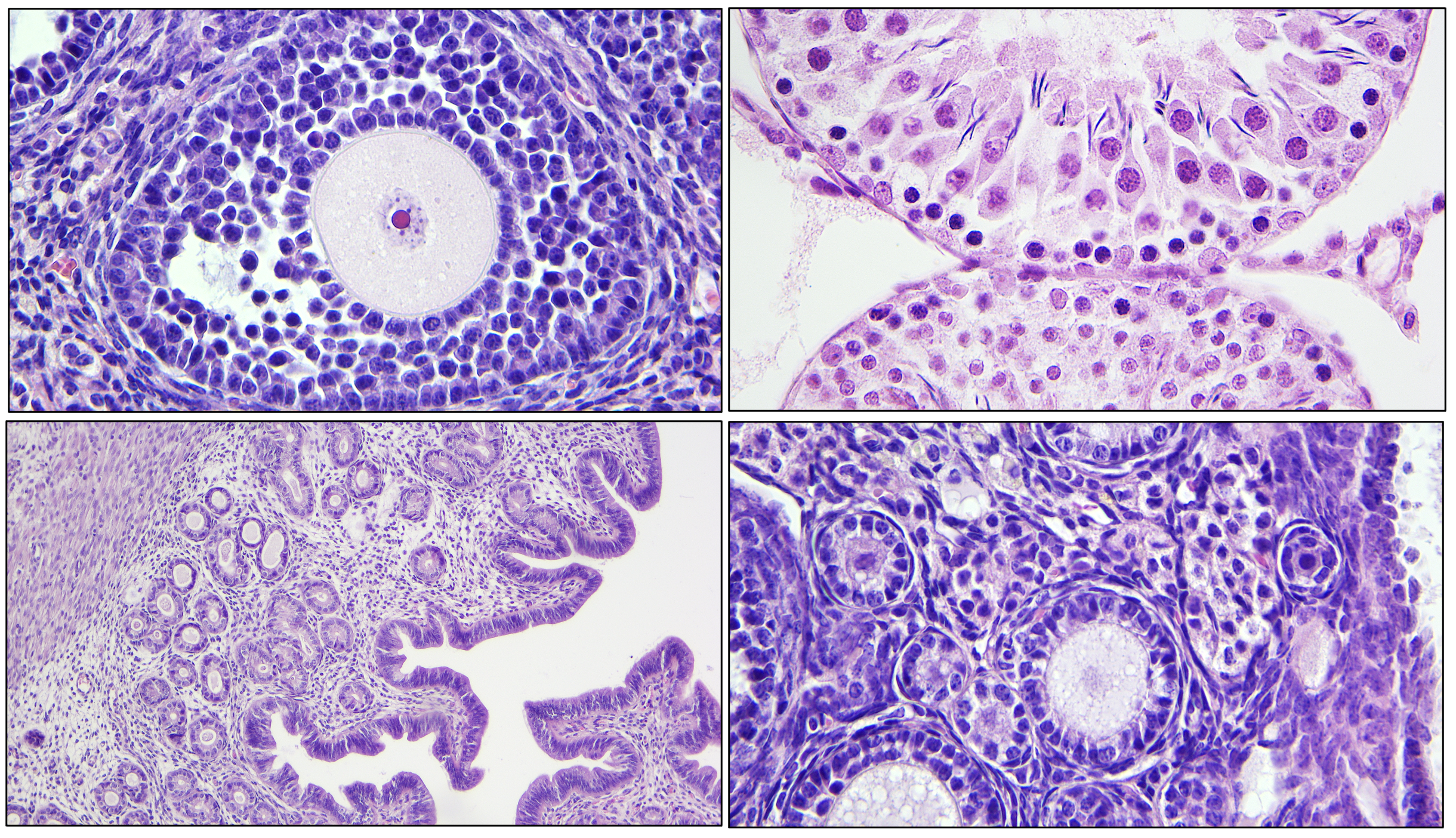
Tissue Processing and Histology
YUCARLAB integrates classical and advanced histological approaches to examine tissue architecture, cellular organization, and pathological remodeling in reproductive and developmental biology. Our laboratory performs tissue fixation, processing, paraffin embedding, microtome sectioning, and histochemical staining to evaluate ovarian, uterine, testicular, and embryonic tissues at the microscopic level. These analyses enable detailed assessment of folliculogenesis, stromal organization, fibrosis, degeneration, inflammatory alterations, and tissue-specific structural changes. In combination with immunohistochemistry and immunofluorescence-based approaches, our histological workflows allow the spatial localization of molecular markers within intact tissue contexts. This integrated tissue-level analysis is essential for linking molecular alterations to cellular morphology, organ architecture, and reproductive function.
- —Paraffin and cryo-embedding techniques
- —Immunohistochemical staining protocols
- —Protein localization in reproductive organs
- —Digital slide scanning and analysis
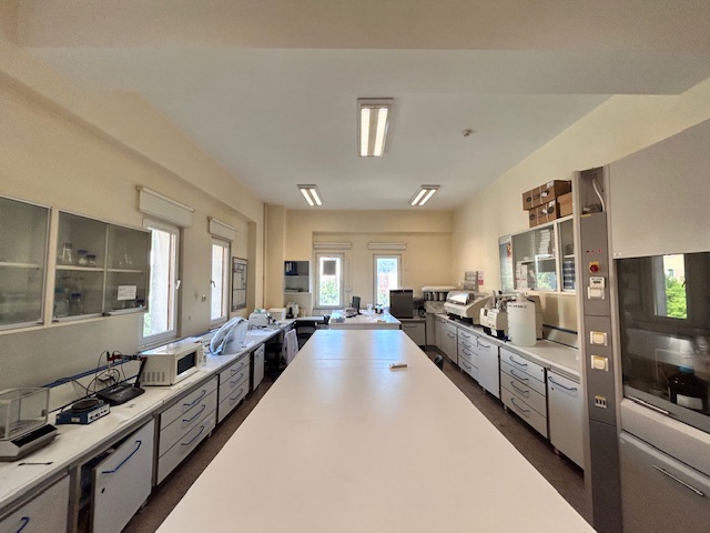
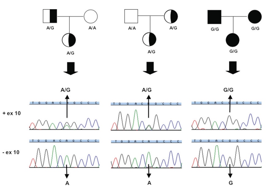
Molecular Biology Laboratory
The molecular biology platform at YUCARLAB is dedicated to elucidating the molecular mechanisms that regulate reproductive physiology, infertility, ovarian aging, fibrosis, oxidative stress, ferroptosis, circadian disruption, Hippo signaling, mTOR activity, and cell fate determination. We employ gene and protein expression-based approaches, including qRT-PCR, immunofluorescence labeling, and molecular marker analysis, to investigate regulatory pathways in cells, embryos, and reproductive tissues. Our research focuses on key molecular targets such as c-Abl, mTOR pathway, Hippo signaling, circadian regulation, mTERT and ferroptosis-associated markers. Through these analyses, we aim to identify molecular signatures associated with reproductive competence, tissue homeostasis, cellular stress responses, and disease-related remodeling.
- —qPCR & RNA sequencing (RNA-seq)
- —Western blot & ELISA assays
- —PCOS and POI molecular pathway analysis
- —Epigenetic modification studies
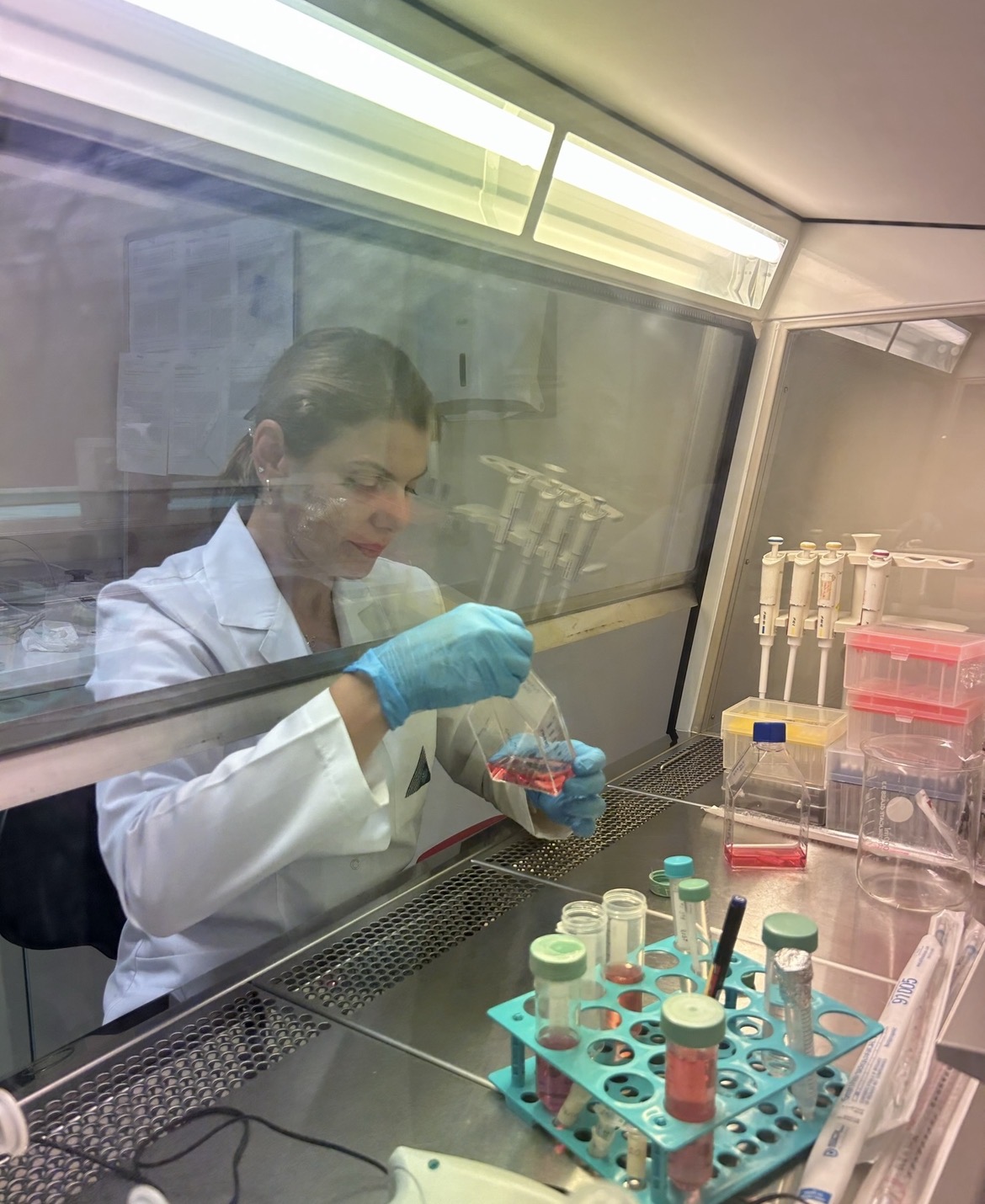
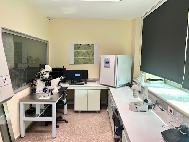
Cell Culture and Image Analysis
YUCARLAB utilizes in vitro cell culture systems and quantitative image analysis to investigate cellular behavior, disease mechanisms, and treatment responses under controlled experimental conditions. Our studies include oocyte and embryo culture, ovarian tissue-based models, germ cell tumor models, and reproductive cell systems designed to evaluate cell viability, proliferation, migration, apoptosis, differentiation, and stress-related molecular responses. High-resolution confocal microscopic images are analyzed using quantitative image-based methods to assess fluorescence intensity, protein localization, nuclear and cytoplasmic distribution, cell number, cell–cell contact, and structural organization. This image-driven analytical approach enables robust, reproducible, and quantitative interpretation of experimental data and supports the identification of cellular and molecular changes underlying reproductive and developmental processes.
- —Fluorescence and confocal microscopy
- —Live cell imaging and time-lapse analysis
- —Automated cell counting and quantification
- —Flow cytometry and cell sorting